
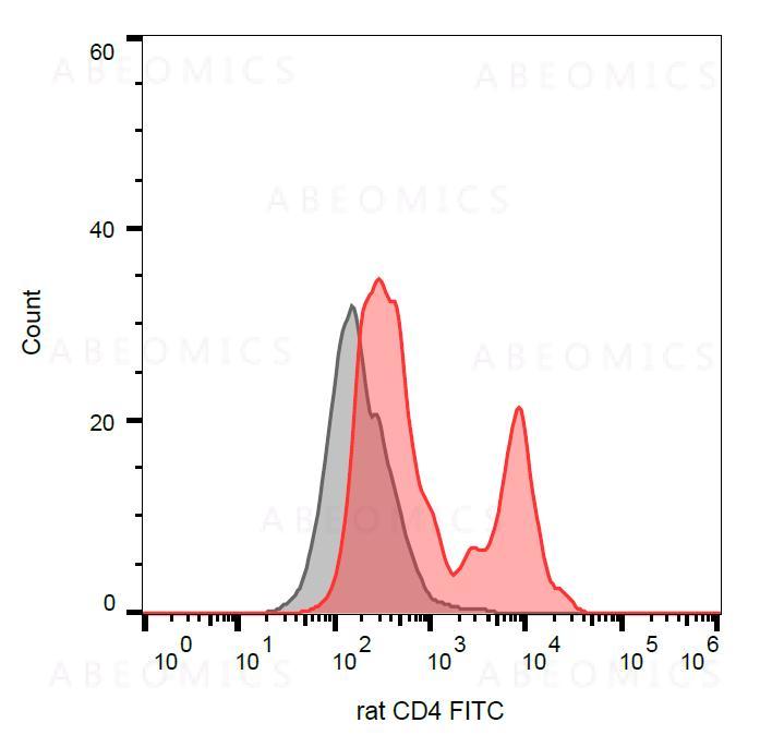
Figure 1: Surface staining of rat thymocytes with anti-rat CD4 (OX-35) FITC.
Conditionnement : 0,1mg
 Téléphone : +1 850 650 7790
View all pathways
View all interactive pathways
Figure 1: Surface staining of rat thymocytes with anti-rat CD4 (OX-35) FITC.
| Amount : | 0.1 mg |
| Isotype : | Mouse IgG2a |
| Storage condition : | Store in the dark at 2-8°C. Do not freeze. Avoid prolonged exposure to light. |
CD4 is a single chain transmembrane glycoprotein of immunoglobulin supergene family. In its extracellular region there are 4 immunoglobulin-like domains (1 Ig-like V-type and 3 Ig-like C2-type). The intracellular region of CD4 associates with p56Lck, a Src-like protein tyrosine kinase. It was described that CD4 segregates into specific detergent-resistant T-cell membrane microdomains. CD4 binds to MHC class II molecules (by CDR2-like region in CD4 domain 1), HIV envelope protein gp120 (by CDR2-like region in CD4 domain 1) and other ligands, such as IL-16 (by to CD4 domain 3) or L-selectin. CD4 is a co-receptor involved in immune response (co-receptor activity in binding to MHC class II molecules) and HIV infection. CD4 regulates T-cell activation, T/B-cell adhesion, T-cell differenciation, T-cell selection and signal transduction. Defects in antigen presentation (MHC class II) cause dysfunction of CD4+ T-cells and their almost complete absence in patients blood, tissue and organs (SCID immunodeficiency).
Flow cytometry: Recommended dilution: 1-4 μg/ml.
For Research Use Only. Not for use in diagnostic/therapeutics procedures.
| Subcellular location: | Cell membrane |
| Post transnational modification: | Phosphorylated by PKC; phosphorylation plays an important role for CD4 internalization. |
| There are currently no product reviews |
