| |
| Gene : | MCAM |
| Gene ID : | 4162 |
| Alternative Name : | MelCAM, MCAM, MUC18,melanoma cell adhesion molecule |
| Immunogen Information : | cultured human umbilical cells |
CD146, also known as MCAM (melanoma cell adhesion molecule) or MUC18, is a heavily glycosylated transmembrane glycoprotein with more than 50% of the mass from carbohydrates. It is expressed on epithelial and endothelial cells, fibroblasts, multipotent mesenchymal stromal cells, activated T cells and activated keratinocytes, and on some cancer cells, especially melanoma. The presence of CD146 on circulating blood cells has been confined to the activated T cells rather than circulating endothelial cells. CD146 mediates heterophilic cell adhesion and regulates monocyte transendothelial migration.
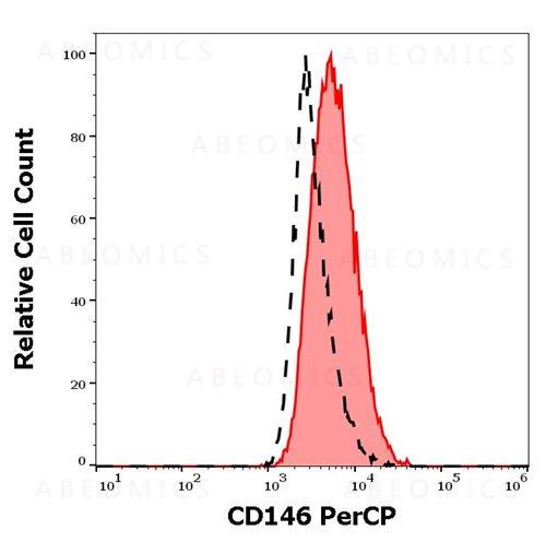
Specificity : The mouse monoclonal antibody P1H12 recognizes an extracellular epitope of CD146, a 118 kDa transmembrane glycoprotein expressed on epithelial and endothelial cells, fibroblasts, multipotent mesenchymal stromal cells, melanoma cells, activated T cells and activated keratinocytes.
Flow cytometry: The reagent is designed for analysis of human blood cells using 10 µl reagent / 100 µl of whole blood or 106 cells in a suspension. The content of a vial (1 ml) is sufficient for 100 tests.
For Research Use Only. Not for use in diagnostic/therapeutics procedures.
| There are currently no product reviews
|