


Application 
| FC |
|---|---|
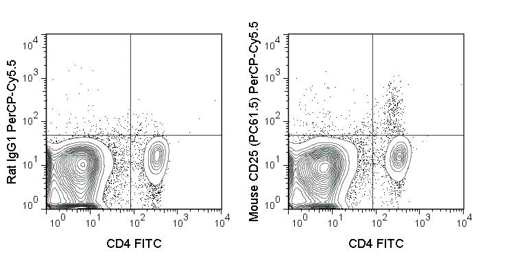
| Isotype | Rat IgG1, lambda |
| Concentration | 0.2 mg/mL |
| Reactivity | Mouse |
| Formulation | 10 mM NaH2PO4, 150 mM NaCl, 0.09% NaN3, 0.1% gelatin, pH7.2 |
| Host | Rat |
| Gene ID | 16184 |
|---|---|
| Gene Name | Il2ra |
| Alternative Name(s) | Interleukin-2 Receptor alpha, IL-2Rα, Ly-43, p55, Tac |
| Format | PerCP-Cy5.5 |
| Preparation | This monoclonal antibody was purified from tissue culture supernatant via affinity chromatography. The purified antibody was conjugated under optimal conditions, with unreacted dye removed from the preparation. It is recommended to store the product undiluted at 4°C, and protected from prolonged exposure to light. Do not freeze. |
| Application Notes | This antibody preparation has been quality-tested for flow cytometry using mouse spleen cells, or an appropriate cell type (where indicated). The amount of antibody required for optimal staining of a cell sample should be determined empirically in your system. |
| Storage Conditions | 2-8°C protected from light |
Liang D, Zuo A, Shao H, Born WK, O’Brian R, Kaplan HJ, and Sun D. 2012. J. Immunol. 188: 5785-5791. (in vivo blocking)
Yu P, Steel JC, Zhang M, Morris JC, Waitz R, Fasso M, Allison JP, and Waldmann TA. 2012. Proc. Natl. Acad. Sci. 109:6187-6192. (in vivo Treg depletion)
Billiard F, Lobry C, Darrasse-Jeze G, Waite J, Liu et al. 2012. Blood. 119: 4656-4664. (in vivo Treg depletion)
Tang S, Moore ML, Grayson JM and Dubey P. 2012. Cancer Res. 72: 1975-1985. (in vivo Treg depletion)
Lee L-F, Logronio K, Tu GH, Zhai W, Ni I, Mei L, Dilley J, Yu J, et al. 2012. Proc. Natl. Acad. Sci. 10.1073. (Flow cytometry).
10F.9G2, J43, PC61 Koehn BH, Ford ML, Ferrer IR, Borom K, Gangappa S, Kirk AD, and Larsen CP. 2008. J. Immunol. 181:5313-5322. (in vivo blocking)
Leithauser F, Meinhardt-Krajina T, Fink K, Wotschke B, Moller P and Reimann J. 2006. Am. J. Pathol. 168(6): 1898-1909. (Immunohistochemistry – frozen tissue)
Hashimoto N, Nabholz M, MacDonald HR, and Zubler RH. 1986. Eur. J. Immunol. 16(3): 317-320. (Blocking)
Ceredig R, Lowenthal JW, Nabholz M, and MacDonald R. 1985. Nature. 314:98-100 (Immunohistochemistry)
Lowenthal JW, Zulber RH, Nabholz M, and MacDonald HR. 1985. Nature. 315(6021): 669-672. (Immunoprecipitation, Blocking)
Provided below are standard protocols that you may find useful for product applications.
Background
The PC61.5 antibody is specific for mouse CD25, a 55 kDa surface protein also known as the Interleukin-2 Receptor alpha chain, or IL-2R alpha. CD25 may bind IL-2 by itself, although with low affinity and without induction of cell signaling. CD25 is also expressed within a high-affinity complex, along with the IL-2R beta chain (CD122) and the common gamma chain (CD132), to form a signaling receptor complex. Expression of CD25 varies during developmental stages of T and B cells, is induced on activated mature T and B cells, and is present on subsets of dendritic cells. CD25 signaling as part of the IL-2 receptor complex triggers T cell activation and proliferation, as well as modulating the differentiation and function of Th17 cells, T regulatory (Treg) cells, and dendritic cells.
The PC61.5 antibody is used as a marker for T cells, B cells and dendritic cell subsets. Expression of CD25, CD4 and the transcription factor Foxp3 is regarded as a phenotypic signature for Treg cells. As such, this antibody is widely used for depletion of Treg cells in vivo, as well as to distinguish Treg cells from naïve or conventional T cells which are CD25-.

