


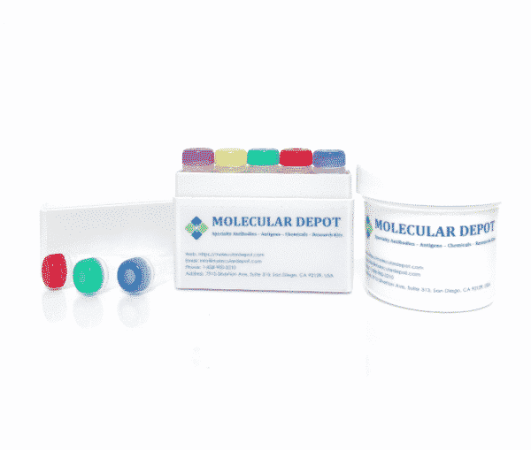
Flow Cytometry Antibody Diluent Buffer
Catalog Number: B2010098 (20 mL)
Flow Cytometry Antibody Diluent Buffer is a ready-to-use, high quality, reproducible solution for the dilution of antibodies used in Flow Cytometry analysis. Custom bulk orders are available upon request.
Product Description
Flow Cytometry Antibody Diluent Buffer
| Catalog number: | B2010098 |
| Lot number: | Batch Dependent |
| Expiration date: | Batch Dependent |
| Volume/Weight | 20 mL |
| pH | 7.4 |
| Supplied as | Ready-to-Use |
| Appearance | Clear Solution |
| Applications | Dilution of antibodies used in Flow Cytometry |
| Shelf-Life | 1 year from date of manufacture |
| Storage: | Keep at 2-8°C |
| Keywords: | Flow Cytometry Antibody Buffer, Antibody Diluent for Flow Cytometry |
| Grade | Biotechnology grade. All components are highly pure (minimum 99%). All solutions are made with Type I ultrapure water (resistivity >18 MΩ-cm) and are filtered on a 0.22 um. |
| References | 1: Ortega-Ferrusola C, Gil MC, Rodríguez-Martínez H, Anel L, Peña FJ, Martín- Muñoz P. Flow cytometry in Spermatology: A bright future ahead. Reprod Domest Anim. 2017 Dec;52(6):921-931. 2: Stewart JJ, Green CL, Jones N, Liang M, Xu Y, Wilkins DE, Moulard M, 3: Wang L, Hoffman RA. Standardization, Calibration, and Control in Flow 4: McFarlin BK. What you see matters: Enhanced detection using image-based flow 5: Vorobjev IA, Barteneva NS. Quantitative Functional Morphology by Imaging Flow 6: Pedersen OH, Nissen PH, Hvas AM. Platelet function investigation by flow 7: Béné MC, Zini G; EHA Scientific Working Group “Diagnosis”, European 8: Go with the immunological flow – Guidelines for flow cytometry. Eur J 9: Rahim A, Meskas J, Drissler S, Yue A, Lorenc A, Laing A, Saran N, White J, 10: Selby LI, Kongkatigumjorn N, Such GK, Johnston AP. HD Flow Cytometry: An 11: Rubbens P, Props R, Boon N, Waegeman W. Flow Cytometric Single-Cell |
| Related Products | Biochemical Buffers and Solutions |
Additional Information
| Weight | 2.6 oz |
|---|---|
| Dimensions | 3.3 × 1.2 × 1.2 in |