

Product Information
- Product Type
- Monoclonal Antibody
- Clone Number
- k1H3
- UniProt No.
- O14933
- NCBI Accession No.
- NP_004214
- Alternative names
- Ubiquitin conjugating enzyme E2 L6, Ubiquitin/ISG15-conjugating enzyme E2 L6, E2 ubiquitin-conjugating enzyme L6, Retinoic acid-induced gene B protein, RIG-B, UbcH8, Ubiquitin carrier protein L6, Ubiquitin-protein ligase L6
Product Specification
- Host
- Mouse
- Reacts With
- Human
- Concentration
- 1mg/ml (determined by BCA assay)
- Formulation
- Liquid in. Phosphate-Buffered Saline (pH 7.4) with 0.02% Sodium Azide, 10% glycerol
- Immunogen
- Recombinant human UBE2L6 (1-152aa) purified from E. coli
- Isotype
- IgG2b kappa
- Purification
- By protein-G affinity chromatography
- Applications
- ELISA, WB, ICC/IF, IHC
- Usage
- The antibody has been tested by ELISA, Western blot, ICC/IF and IHC analysis to assure specificity and reactivity. Since application varies, however, each investigation should be titrated by the reagent to obtain optimal results.
- Storage
- Can be stored at +2C to +8C for 1 week. For long term storage, aliquot and store at -20C to -80C. Avoid repeated freezing and thawing cycles.
Data
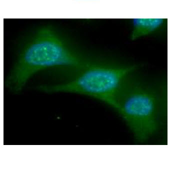
Immunocytochemistry/Immunofluorescence (ICC/IF)
ICC/IF analysis ofUBE2L6 in HeLa cells. The cell was stained with AUB0907 (1:100). The secondary antibody (green) was used Alexa Fluor 488. DAPI was stained the cell nucleus (blue).
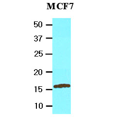
Western blot analysis (WB)
The cell lysate of MCF7 (20ug) was resolved by SDS-PAGE, transferred to NC membrane and probed with anti-human UBE2L6 (1:2000). Proteins were visualized using a goat anti-mouse secondary antibody conjugated to HRP and an ECL detection system.
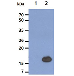
The cell lysates (5ug) were resolved by SDS-PAGE, transferred to PVDF membrane and probed with anti-human UBE2L6 antibody (1:2000). Proteins were visualized using a goat anti-mouse secondary antibody conjugated to HRP and an ECL detection system.
Lane 1.: 293T cell lysate
Lane 2.: UBE2L6 Transfected 293T cell lysate
Lane 1.: 293T cell lysate
Lane 2.: UBE2L6 Transfected 293T cell lysate
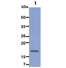
The cell lysate (20ug) was resolved by SDS-PAGE, transferred to PVDF membrane and probed with anti-human UBE2L6 antibody (1:2000). Proteins were visualized using a goat anti-mouse secondary antibody conjugated to HRP and an ECL detection system.
Lane 1.: Jurkat cell lysate
Lane 1.: Jurkat cell lysate
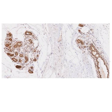
Immunohistochemistry (IHC)
Paraffin embedded sections of human breast lobule tissue were incubated with anti-human UBE2L6 (1:50) for 2 hours at room temperature. Antigen retrieval was performed in 0.1M sodium citrate buffer and detected using Diaminobenzidine (DAB)
Note: For research use only. This product is not intended or approved for human, diagnostics or veterinary use.