

Monoclonal Antibody to Fibrillarin/Nop1p (Clone: 38F3)
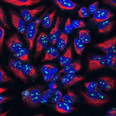
Figure-1: High magnification confocal image of HeLa cells stained with fibrillarin antibody (34-1031), dilution 1:100 in green, and costained with(34-1126), chicken polyclonal antibody to vimentin, in red, 1:10,000. Nuclear DNA is revealed with the DAPI stain in blue. The fibrillarin antibody shows strong staining of nucleoli in the nucleus, while the vimentin antibody reveals cytoplasmic intermediate filaments.
