Calcein AM Cell Viability Assay Kit (1000 assays)

Cat# 30026
Size : 1kit
Brand : Biotium
Contact local distributor :

Phone : +1 850 650 7790
| Apoptosis/viability marker | Metabolic activity, Live cell stain |
|---|---|
| For live or fixed cells | For live/intact cells |
| Detection method/readout | Microplate reader (fluorescence), Fluorescence microscopy, Live cell imaging, Flow cytometry |
| Assay type/options | Endpoint assay, Short term staining (<24h) |
| Colors | Green |
| Excitation/Emission | 485/530 nm (end product) |
| Storage Conditions | Store at -10 to -35 °C, Protect from light, Desiccate |
Product Description
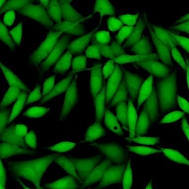
The Calcein AM Cell Viability Assay Kit is designed to quantify live cell numbers based on their endogenous esterase activity and plasma membrane integrity.
- Quantitate live cells using green fluorescence
- 30-min assay
- True endpoint viability assay; only live cells retain signal
- For fluorescence microplate reader, fluorescence microscopy, or flow cytometry
- Ex/Em 485/530 nm
Calcein AM is a widely used green fluorescent cell marker. Calcein AM is itself non-fluorescent and membrane-permeant, and thus can be introduced into cells via incubation. Once inside the cells, endogenous esterases hydrolyze the compound into the highly negatively charged green fluorescent dye calcein, which is retained in the cytoplasm in live cells. Only viable cells with intact plasma membranes retain fluorescence, making this a true end-point assay for cell viability. The fluorescent signal generated from the assay is proportional to the number of living cells in the sample. Calcein AM is also a useful tool for cell tracing.
We supply Calcein AM in a variety of packaging sizes and formats. See our full line of Cell Viability & Apoptosis Assays.
References
1. Brain Res (2009) 1275, 87-95. doi: 10.1016/j.brainres.2009.04.008
2. J Hepatol (2010) 52(5), 690-697. doi: 10.1016/j.jhep.2009.12.025
3. PLoS ONE (2011) 6(6), e20301. doi:10.1371/journal.pone.0020301
4. PLoS ONE (2012) 7(4), e35007. doi:10.1371/journal.pone.0035007
5. Cancer Biol Ther (2013) 14(4),357-364.DOI: 10.4161/cbt.23623
6. Toxics (2014) 2, 258-275. doi:10.3390/toxics2020258
7. Cell Physiol Biochem (2015) 36, 384-394. DOI: 10.1159/000430257
8. RSC Adv (2015) 5, 47749-47756. https://doi.org/10.1039/C5RA06861D
9. Biomaterials (2018) 179, 60-70. doi:10.1016/j.biomaterials.2018.06.027
10. Sci Rep (2018) 8, 766. https://doi.org/10.1038/s41598-017-17539-z
11. Mol Cancer Ther (2019) 18(6), 1092. DOI: 10.1158/1535-7163.MCT-18-1313
12. Sci Rep (2019) 9, 2147. https://doi.org/10.1038/s41598-019-38590-y
 Powered by Bioz
 See more details on Bioz