

Monoclonal Antibody to a-internexin/NF66 (Clone: 1D2)
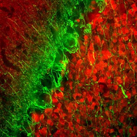
Figure-1: Immunofluorescent analysis of rat cerebellum section stained with mouse mAb to α-internexin,(34-1052), dilution 1:5,000 in green, and costained with chicken pAb to calretinin, 1:2,000 in red. Following transcardial perfusion of rat with 4% paraformaldehyde, brain was post fixed for 24 hours, cut to 45μM, and free-floating sections were stained with the above antibodies. The α-internexin antibody selectively stains neuronal processes, in particular parallel fibers, the axons of granule cells. Calretinin antibody stains interneurons predominantly in the molecular layer of the cerebellum.
