NuSep
 |
||
NuSep offers a complete range of precast gels for protein electrophoresis.
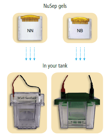
Compatible with all 10 cm tanks
3 different types of gel to fit all tanks :
 2 minutes visualization by UV
All nUView precast gels incorporate a unique formulation allowing protein bands to be visualized in only 2 minutes in the presence of ultraviolet (UV) light. More information
Flexibility to run in different buffer systems
NuSep gel can be developed in a range of Tris buffers with different counter ions, each producing subtle variation to the migration pattern. Compatible with Tris-Glycine running buffers 
Website : www.nusep.com
| ||
Protein Electrophoresis
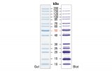
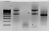
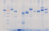
Protein electrophoresis is a gel electrophoresis technique that allows the analysis of a mixture of proteins. The gel used is composed of acrylamide, which polymerises to form a mesh network. Gel electrophoresis allows proteins to be separated for analysis. It relies on the ability of charged proteins to migrate through the mesh of a gel when an electric current is applied. The analysis of a mixture by gel electrophoresis is carried out on so-called denatured proteins, i.e. proteins that have lost their tertiary and secondary structures. An anionic detergent covers the denatured polymer chains, giving them a charge (constant charge/amino acid ratio). The individual proteins in a complex mixture are thus covered with a "coat" of negative charges. When a continuous voltage is applied between the ends of a gel where the complex mixture of proteins has been deposited, the proteins migrate through the meshes constituting the gel. The gel mesh will hold less of the lower molecular weight molecules which will then have the greatest migration. Longer molecules will be retained more between the gel meshes and will have a lower relative migration. The definition of the separation can be increased or decreased by using a more or less loosely meshed gel. A molecular weight marker is used during migration to give a molecular weight scale that will allow the size of each protein in the mixture to be determined.
In order to visualise the proteins after migration, a specific stain is most often used. The most commonly used stain is Coomassie blue but it is also possible to perform a silver stain. A molecular weight marker is used during migration to give a molecular weight scale that will allow the size of each protein in the mixture to be determined.