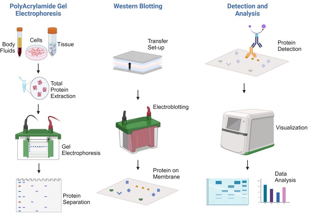
Experimental Protocol for Western Blotting
Western blot is a technique used to identify specific proteins from a complex mixture of proteins. Prior to Western blot, samples undergo a size-based protein separation step through gel electrophoresis. Western blot includes the transfer of size-based separated proteins onto a solid support and detection of specific proteins within the sample. Western blots are both highly sensitive and specific; the process incorporates a highly selective antibody detection system to identify target proteins within a sample which allows the researcher to accurately detect proteins based on the molecular weights.
Individual steps in the procedure are modifiable and factors like gel concentration, duration of incubation, and voltage used in the run time can be optimized depending on the needs. These attributes make Western blot a highly accurate, flexible, and semi-quantitative method for identifying proteins.

Protein Separation by Gel Electrophoresis
Before Western blot, the proteins within a sample must be separated based on their size. Typically, this is done using SDS-PAGE (Sodium Dodecyl-Sulfate Poly-Acrylamide Gel Electrophoresis), wherein the concentration of polyacrylamide along with the buffer system influences the mobility of the proteins through the gel depending on their molecular weight. The gel in SDS-PAGE has two distinct layers, termed the “stacking” gel (top layer) and “separating” or “resolving” gel (bottom layer).
As depicted in the table below, the resolving gel concentration should correspond to the size of your target protein.
| Protein size (kDa) | Polyacrylamide concentration |
| ≥500 | 5% |
| 25-200 | 8% |
| 15-100 | 10% |
| 10-70 | 12.5% |
| 12-45 | 15% |
| 4-40 | 20% |
SDS-PAGE gels can be purchased, or easily made in-house. A common recipe for making an SDS-PAGE gel is as follows. If using pre-made gel, skip to the next section.
| Reagents | 6% Stacking gel | 10% Separating gel |
| ddH2O | 2 mL | 3 mL |
| 1 M Tris-HCl | 400 uL pH 6.7 | 2.1 mL pH 8.9 |
| 30% Acrylamide/Bis- Acrylamide | 600 uL | 2.8 mL |
| 10% SDS | 36 uL | 80 uL |
| 10% APS (Ammonium Persulfate) | 24 uL | 50 uL |
| TEMED (TetraMethylEthyleneDiamine) | 4 uL | 6 uL |
- Assemble the gel casting kit per the manufacturer instructions. This includes inserting the glass plates into the casting frame and locking them in place.
- Insert the casting frame into the casting stand. Place the comb appropriately.
- Using a marker, mark the glass ~ 1cm underneath the comb. This indicates the level of separating gel needed.
- Remove the comb. Using a pipette, add a small amount of water into the gel chamber to ensure no leaks are present. Remove the water with a KimWipe.
- Mix together all the ingredients in the separating gel, making sure to add TEMED last.
- Pipet the separating gel into the chamber, up to your marker.
- To remove bubbles, gently pipet a small amount of isopropyl alcohol (IPS) onto the top of the gel. Allow the gel to set for 30 minutes.
- Remove the IPA by gently inserting a KimWipe into the chamber.
- Mix together all the ingredients in the stacking gel, making sure to add TEMED last.
- Pipet the stacking gel into the chamber, up to the top of the glass plate.
- Insert your comb. Allow the gel to set for another 30 minutes.
Sample Preparation
Proteins are typically extracted from cells or tissues using various extraction buffers containing detergents, salts, and protease inhibitors to maintain protein integrity and stability.
The amount of protein present in the sample is estimated by following methods:
- Direct method based on absorption at 280nm by UV-spectroscopy.
- Bradford Protein Assay (Bradford Assay Calculator)
- BCA Protein Quantification Assay (Amplite® Colorimetric or Amplite® Fluorimetric or Portelite™ Fluorimetric Assay kits).
To denature proteins and disrupt any non-covalent interactions, a reducing agent such as β-mercaptoethanol or dithiothreitol (DTT) is added to the sample buffer. Additionally, heating the samples at around 95°C for 5 minutes or at 70°C for 10 min helps to fully denature the proteins. A loading dye is added to the protein samples to increase their density, provide color for visualization, and track the migration of the samples during electrophoresis. The dye typically contains a tracking dye (e.g., bromophenol blue or xylene cyanol) and a dense agent (e.g., glycerol) to help the samples sink into the wells.
Gel Electrophoresis
Equipment needed :
-
Electrophoresis chamber and power supplies
- Micropipette
Reagents needed :
- SDS-PAGE gel
- 1X Running buffer (Recipe for 10X Running buffer)
- Protein Samples
- Protein Ladder (PageTell™ Prestained 10-250 kDa Ladder or ProLite™ 5-245 kD Protein Ladder)
- Insert the gel cassette into the electrode chamber. Place the electrode chamber into the electrophoresis tank.
- Remove the comb on the gel.
- Add ~200 mL of 1x running buffer to the inner electrode chamber.
- Fill the outer chamber of the tank to the indicator mark present on the outside of the tank. Make sure the running buffer covers the gel completely.
- Heat samples in a water bath at 95°C for 5 min or at 70°C for 10 min.
- Add your samples and protein ladders to identified wells. The volume depends on the sample, but is generally between 5 - 15 uL per well. The volume in each lane should be made equal by using ddH20 or lysis buffer.
- Connect the electrophoresis tank to your power supply, red (+) to red, black (-) to black.
- Run your gel for the desired voltage and for the desired time. Generally, these parameters are 100 - 150 V for 40 to 60 minutes.
The protein separated on the gel may be stained with ProLite™ Orange Protein Gel Stain for visualizing total protein present on the gel.
Electrotransfer
Next, the separated proteins present on the gel are transferred onto a membrane through a process called electrotransfer (also electroblotting).
Equipment needed :
- Transfer apparatus
- Sponge or foam, cut to the same dimension of the gel
- 6 filter papers, cut to the same dimension of the gel
- Assembly tray or blotting cassette
Reagents needed :
- Ice
- Methanol
- PVDF (polyvinylidene fluoride) or Nitrocellulose membrane, cut to the same dimensions of the gel
- 1X Transfer buffer (Recipe for 20X Transfer buffer)
- Wet the sponge, filter papers, and membrane with methanol.
- Carefully retrieve the gel from the gel cassette.
- Carefully assemble an electro transfer sandwich by stacking in the following order :
-
(a) Assembly tray bottom
-
(b) Pressure sponge
-
(c) Filter papers
-
(d) Gel
-
(e) PVDF membrane
-
(f) Filter papers
-
(g) Assembly tray top
-
- Ensure no air bubbles are present in the sandwich. Gently squeeze out extra liquid.
- Position the sandwich into the electroblotting tank, on ice.
- Add transfer buffer to the electroblotting tank, covering the sandwich completely.
- Connect the electroblotting tank to your power supply, red (+) to red, black (-) to black.
- Run the electroblotting system for the desired duration, which ranges between 45 - 90 minutes.
Blocking
A blocking step will prevent antibodies from nonspecifically binding to the membrane. Blocking removes potential background noise from the membrane, simplifying protein analysis. Commonly used blockers are 2-5% BSA or non-fat dry milk in buffered saline solution (TBS or PBS).
Table1. Comparison of blocking buffers for western blotting.
| Blocking Buffer | Benefits | Considerations |
| Skim milk |
|
Contains biotin and phosphoproteins, which can interfere with streptavidin-biotin detection strategies and detection of phosphorylated target proteins. Due to the number of proteins within milk, milk may mask some antigens and lower the detection limit of the western blot. |
| Purified proteins |
|
More expensive than taraditional non-fat milk formulations. |
| Bovine serum albumin (BSA) |
|
Various grades of BSA are commercially available that can impacte signal-to-noise. BSA is generally a weaker blicker, which can result in more non--specific antibody binding, but can increase the detection sensitivity for low-abundant proteins. |
Primary and Secondary Antibody Incubation
The protein of interest in a Western blot can be detected by using either a labeled primary antibody or an unlabeled primary antibody + labeled secondary antibody.
- Add the primary antibody in 5% BSA (bovine serum albumin) to the membrane.
- Incubate the membrane overnight at 4°C on a shaker or for 1 hour at room temperature.
- If using a secondary antibody, wash the membrane thrice with TBST. Incubate the membrane with the secondary antibody for 1 hour at room temperature.
- Wash the membrane three times with TBST.
Detection
In Western blotting, detection methods are used to visualize and quantify the presence of specific proteins that have been transferred from the gel to the membrane. Several detection methods can be employed, depending on the specific requirements of the experiment and the type of label present in your detection antibody. Here are some commonly used detection methods in Western blotting:
Chemiluminescence
Chemiluminescent substrates like Amplite® West ECL HRP Substrate generate light upon enzymatic reaction with horseradish peroxidase (HRP) or alkaline phosphatase (AP), which are conjugated to secondary antibodies. It is a sensitive method and gives a long-lasting signal.
Fluorescence
Fluorescently labeled secondary antibodies directly bind to primary antibodies, or primary antibodies are detected using fluorophore-conjugated secondary antibodies. This approach allows for multiplexing capabilities, high sensitivity, and compatibility with various imaging systems.
The Antibody and Protein Labeling Selection Guide can be helpful in planning Western blotting and other immunoassays by providing information on choosing the most suitable antibodies for specific experimental needs.
Colorimetric Detection
Enzymatic substrates produce a colored reaction product upon reaction with HRP or AP conjugated to secondary antibodies which can be utilized for visualizing protein in western blot.
Analysis
In Western blot analysis, clear blot images are imaged using specialized equipment. Band intensity can be quantified with image analysis software, often normalizing to internal controls for accurate comparisons. Statistical tests assess differences between the samples analyzed, helping draw meaningful conclusions.
Explore our Digital Catalog on Western Blotting Assays, meticulously curated to present a diverse range of products, categorized for ease of navigation and discover solutions tailored to your needs.

