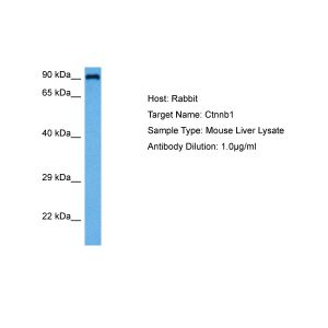
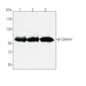
CTNNB1 Antibody (Phospho-Tyr654)

Referencia OASG01072
embalaje : 100ul
Marca : Aviva Systems Biology
Contact local distributor :
 Teléfono : +1 850 650 7790
CTNNB1 Antibody (Phospho-Tyr654) (OASG01072)
| Datasheets/Manuals | Printable datasheet for anti-CTNNB1 (OASG01072) antibody |
|---|
| Predicted Species Reactivity | Human, Mouse, Rat |
|---|---|
| Product Format | Liquid. PBS containing 50% glycerol, 0.5% BSA and 0.02% sodium azide. |
| Clonality | Polyclonal |
| Isotype | IgG |
| Host | Rabbit |
| Application | ELISA, IF, IHC, WB |
| Additional Information | Cellular Location: Cytoplasm Nucleus Cytoplasm, cytoskeleton Cell junction, adherens junction Cell junction Cell membrane Cytoplasm, cytoskeleton, microtubule organizing center, centrosome. Cytoplasm, cytoskeleton, spindle pole. Cell junction, synapse Note=Colocalized with RAPGEF2 and TJP1 at cell-cell contacts (By similarity). Cytoplasmic when it is unstabilized (high level of phosphorylation) or bound to CDH1. Translocates to the nucleus when it is stabilized (low level of phosphorylation). Interaction with GLIS2 and MUC1 promotes nuclear translocation. Interaction with EMD inhibits nuclear localization. The majority of beta-catenin is localized to the cell membrane. In interphase, colocalizes with CROCC between CEP250 puncta at the proximal end of centrioles, and this localization is dependent on CROCC and CEP250. In mitosis, when NEK2 activity increases, it localizes to centrosomes at spindle poles independent of CROCC. Colocalizes with CDK5 in the cell-cell contacts and plasma membrane of undifferentiated and differentiated neuroblastoma cells. Interaction with FAM53B promotes translocation to the nucleus. |
| :: | Tissue Specificity: Expressed in several hair follicle cell types: basal and peripheral matrix cells, and cells of the outer and inner root sheaths. Expressed in colon. Present in cortical neurons (at protein level). |
| Reconstitution and Storage | Store at -20°C. Avoid repeated freeze/thaw cycles. |
| Immunogen | Synthesized peptide derived from human Catenin-Beta around the phosphorylation site of Y654. Location: 590-670aa. |
| Purification | The antibody was affinity-purified from rabbit antiserum by affinity-chromatography using epitope-specific immunogen. |
| Concentration | 1 mg/ml |
| Target Post-Translational Modification | Phosphorylation at Ser-552 by AMPK promotes stabilizion of the protein, enhancing TCF/LEF-mediated transcription . Phosphorylation by GSK3B requires prior phosphorylation of Ser-45 by another kinase. Phosphorylation proceeds then from Thr-41 to Ser-37 and Ser-33. Phosphorylated by NEK2. EGF stimulates tyrosine phosphorylation. Phosphorylation on Tyr-654 decreases CDH1 binding and enhances TBP binding. Phosphorylated on Ser-33 and Ser-37 by HIPK2 and GSK3B, this phosphorylation triggers proteasomal degradation . Phosphorylation on Ser-191 and Ser-246 by CDK5. Phosphorylation by CDK2 regulates insulin internalization. Phosphorylation by PTK6 at Tyr-64, Tyr-142, Tyr-331 and/or Tyr-333 with the predominant site at Tyr-64 is not essential for inhibition of transcriptional activity. Ubiquitinated by the SCF(BTRC) E3 ligase complex when phosphorylated by GSK3B, leading to its degradation. Ubiquitinated by a E3 ubiquitin ligase complex containing UBE2D1, SIAH1, CACYBP/SIP, SKP1, APC and TBL1X, leading to its subsequent proteasomal degradation . S-nitrosylation at Cys-619 within adherens junctions promotes VEGF-induced, NO-dependent endothelial cell permeability by disrupting interaction with E-cadherin, thus mediating disassembly adherens junctions. O-glycosylation at Ser-23 decreases nuclear localization and transcriptional activity, and increases localization to the plasma membrane and interaction with E-cadherin CDH1. Deacetylated at Lys-49 by SIRT1. |
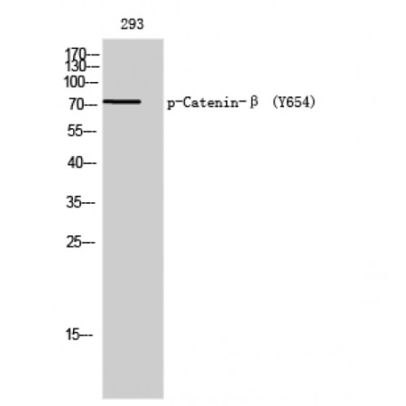
| Specificity | Phospho-Catenin-Beta (Tyr654) Polyclonal Antibody detects endogenous levels of Catenin-Beta protein only when phosphorylated at Y654. |
| Application Info | WB: 1:500~2000 IHC: 1:100~300 ELISA: 1:40000 Optimal dilutions should be determined by the end user. |
| Gene Symbol | CTNNB1 |
|---|---|
| Gene Full Name | catenin (cadherin-associated protein), beta 1, 88kDa |
| Alias Symbols | EVR7, CTNNB, MRD19, NEDSDV, armadillo |
| NCBI Gene Id | 1499; 12387; 84353 |
| Description of Target | Key downstream component of the canonical Wnt signaling pathway. In the absence of Wnt, forms a complex with AXIN1, AXIN2, APC, CSNK1A1 and GSK3B that promotes phosphorylation on N-terminal Ser and Thr residues and ubiquitination of CTNNB1 via BTRC and its subsequent degradation by the proteasome. In the presence of Wnt ligand, CTNNB1 is not ubiquitinated and accumulates in the nucleus, where it acts as a coactivator for transcription factors of the TCF/LEF family, leading to activate Wnt responsive genes. Involved in the regulation of cell adhesion, as component of an E-cadherin:catenin adhesion complex. Acts as a negative regulator of centrosome cohesion. Involved in the CDK2/PTPN6/CTNNB1/CEACAM1 pathway of insulin internalization. Blocks anoikis of malignant kidney and intestinal epithelial cells and promotes their anchorage-independent growth by down-regulating DAPK2. Disrupts PML function and PML-NB formation by inhibiting RANBP2-mediated sumoylation of PML . Promotes neurogenesis by maintaining sympathetic neuroblasts within the cell cycle. |
| Uniprot ID | P35222, Q02248, Q9WU82 |
| Molecular Weight | 75 kDa |