Goat anti-SGLT1

Referencia EB09310
embalaje : 100ug
Marca : Everest Biotech
Contact local distributor :
 Teléfono : +1 850 650 7790
| Code | Name | Applications | Availability | Size | Tested species | Grade |
|---|---|---|---|---|---|---|
| EB09310 | Goat Anti-SGLT1 Antibody | Pep-ELISA, WB, ICC, IHC | Contact sales for delivery estimate | 100µg specific antibody in 200µl | Human, Mouse |
Target Protein
Principal Names: SLC5A1, solute carrier family 5 (sodium/glucose cotransporter), member 1, D22S675, NAGT, SGLT1, Human Na+/glucose cotransporter 1 mRNA, complete cds, sodium/glucose cotransporter 1, solute carrier family 5 (sodium/glucose transporter), member 1Official Symbol: SLC5A1
Accession Number(s): NP_000334.1
Human GeneID(s): 6523
Immunogen
Peptide with sequence C-KETIEIETQVPEKKK, from the internal region of the protein sequence according to NP_000334.1.
Please note the peptide is available for sale.
Purification and Storage
Purified from goat serum by ammonium sulphate precipitation followed by antigen affinity chromatography using the immunizing peptide.
Supplied at 0.5 mg/ml in Tris saline, 0.02% sodium azide, pH7.3 with 0.5% bovine serum albumin.
Aliquot and store at -20°C. Minimize freezing and thawing.
Applications Tested
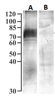
Peptide ELISA: antibody detection limit dilution 1:4000.Western blot: Approx 75kDa band observed in isolated Brush Border membranes from Human Kidney (calculated MW of 73.5kDa according to NP_000334.1). Recommended concentration: 0.3-1µg/ml. Primary incubation was 1 hour. This antibody has been successfully used in WB on Mouse in the following papers, PMID: 34198013, 30503674 and 28174043.
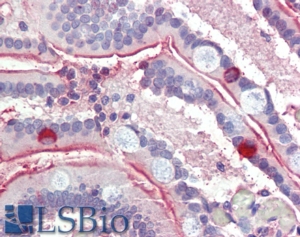
IHC: In paraffin embedded Human Small Intestine shows staining of the glycocalix. Recommended concentration: 3-6µg/ml. This antibody has been successfully used in IHC on Mouse in the following paper, PMID: 34198013.
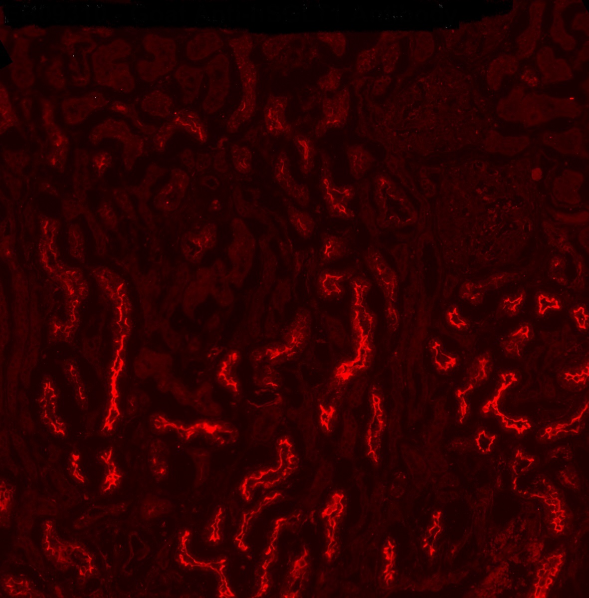
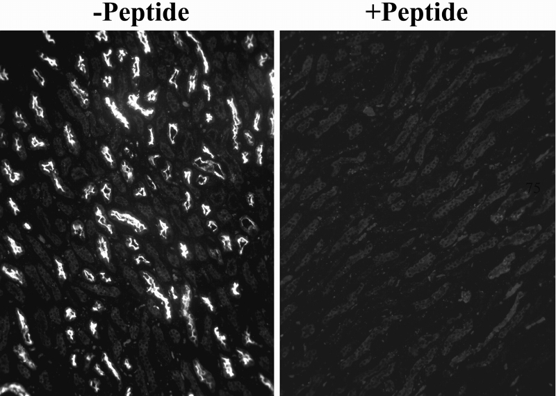
Immunocytochemistry: Fig A.Frozen sections of Human Kidney show staining of the brush borders in DCT, while only weak background was observed in Rat and Pig Kidney. Recommended concentration: 2-4ug/ml. Fig B.PFA-fixed 4µm cryosections of Human Kidney shows staining of the brush border of proximal straight tubules (S3 segments) in the outer stripe zone. The signal drops to background levels upon blocking with the immunizing peptide.
Species Reactivity
Tested: Human, MouseExpected from sequence similarity: Human
Specific References
This antibody has been successfully used in Western blot and IHC on Mouse:
Barun Das, Kevin Okamoto, John Rabalais, Jocelyn A. Young, Kim E. Barrett, and Mamata Sivagnanam
Aberrant Epithelial Differentiation Contributes to Pathogenesis in a Murine Model of Congenital Tufting Enteropathy.
Cell Mol Gastroenterol Hepatol. 2021; 12(4): 1353-1371.
PMID: 34198013
This antibody has been successfully used in Western blot on Mouse:
Yui Yamazaki, Kyoko Arita, Shinichi Harada, Shogo Tokuyama
Activation of c-Jun N-terminal kinase and p38 after cerebral ischemia upregulates cerebral sodium-glucose transporter type 1
Journal of Pharmacological Sciences (November 2018)
PMID: 30503674
This antibody has been successfully used in Western blot on Mouse:
Yamazaki Y, Harada S, Wada T, Hagiwara T, Yoshida S, Tokuyama S
Sodium influx through cerebral sodium-glucose transporter type 1 exacerbates the development of cerebral ischemic neuronal damage.
Eur J Pharmacol. 2017 Mar 15;799:103-110.
PMID: 28174043
- Western blot Guidelines
- Everest Western blot labeling
- Western blot Trouble shooting guide
- IHC staining on paraffin sections
- Immunofluorescence Protocol
- Flow Cytometry Protocol
- MSDS - All Goat antibodies
- Tissue Lysate Preparation
- Cell Lysate Preparation
- Blocking with the immunizing peptide
- LsBio IHC Protocol