VEA, very early activation antigen, AIM, EA1, MLR3, gp34/28, Leu-23
Description
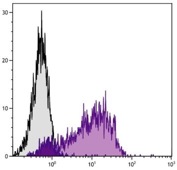
CD69, also known as very early activation (VEA) antigen, is a disulfide-linked transmembrane homodimer whose differentially glycosylated subunits range from 35-39 kDa. It is a C-type lectin most closely related to the NKR-P1 and Ly-49 NK cell-activation molecules. CD69 is widely expressed on hematopoietic cells, including lymphocytes, neutrophils and eosinophils. Although not detectable on resting lymphocytes, its expression is rapidly upregulated upon activation of T, B and NK cells, and neutrophils. Constitutive expression of CD69 on subsets of thymocytes suggests that it may be involved in regulation of developmental events in addition to its role in activation of a variety of hematopoietic cells. The monoclonal antibody H1.2F3 augments PMA-induced T-cell proliferation and induces redirected lysis of Fc receptor-bearing target cells by NK cells.
Immunogen
Mouse dendritic epidermal cell line Y245
Conjugate
PE (R-phycoerythrin)
Buffer Formulation
Phosphate buffered saline containing < 0.1% sodium azide and a stabilizer
1. Yokoyama WM, Koning F, Kehn PJ, Pereira GM, Stingl G, Coligan JE, et al. Characterization of a cell surface-expressed disulfide-linked dimer involved in murine T cell activation. J Immunol. 1988;141:369-76. (Immunogen, FC, IP, Costim)
2. Karlhofer FM, Yokoyama WM. Stimulation of murine natural killer (NK) cells by a monoclonal antibody specific for the NK1.1 antigen. IL-2-activated NK cells possess additional specific stimulation pathways. J Immunol. 1991;146:3662-73. (FC, Activ)
3. Podd BS, Thoits J, Whitley N, Cheng H, Kudla KL, Taniguchi H, et al. T cells in cryptopatch aggregates share TCR γ variable region junctional sequences with γδ T cells in the small intestinal epithelium of mice. J Immunol. 2006;176:6532-42. (IHC-FS)

 Teléfono : +1 850 650 7790