

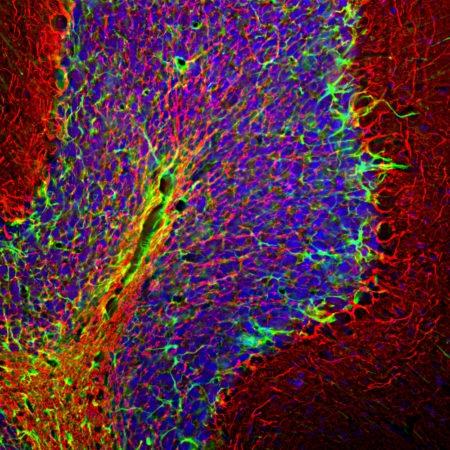
Figure-1: Immunohistological analysis of a rat cerebellum section stained with chicken pAb to NF-H, (34-1080), dilution 1:5,000 in red, and costained with rabbit pAb to GFAP,(34-1042), dilution 1:5,000 in green. The blue is DAPI staining of nuclear DNA. Following transcardial perfusion with 4% paraformaldehyde, brain was post fixed for 24 hours, cut to 45μM, and free floating sections were stained with above antibodies. The NF-H antibody labels network of axons of different neurons, while the GFAP antibody stains astrocytes and other glial cells.
| Format : | Conc. IgY prep. |
| Amount : | 50 µl |
| Isotype : | Chicken, IgY |
| Content : | Antibody is supplied as an aliquot of concentrated IgY prep. |
| Storage condition : | Store the antibody at 4°C; stable for 6 months. For long-term storage; store at -20°C. Avoid repeated freeze and thaw cycles. |
WB: 1:20,000-1:50,000. IF/ICC, IHC: 1:20,000.
For Research Use Only. Not for use in diagnostic/therapeutics procedures.
| Subcellular location: | Cytoplasm |
| Post transnational modification: | Phosphorylated in the head and rod regions by the PKC kinase PKN1, leading to the inhibition of polymerization. |
| BioGrid: | 110819. 25 interactions. |
| There are currently no product reviews |