AgriseraECL Bright
Referentie AS16ECL-N-100
Formaat : 2x50ml
Merk : Agrisera
AS16 ECL-N-100 | low pico to mid femtogram detection
- Product Info
-
Quantity: 2 x 50 ml, two component ready to use solutions, enough for 50 midi blots (6.8 x 8.1 cm) AS16 ECL-N-10, AgriseraECL Bright 10 ml (5 ml of component A + 5 ml of component B, trial pack)AS16 ECL-N-100, AgriseraECL Bright 100 ml (50 ml of component A + 50 ml of component B)AS16 ECL-N-1L, AgriseraECL Bright 1L (0.5 L of component A + 0.5 L of component B) Storage: Store at 2°C to 8°C.Mixed working reagent is stable for several days at room temperature or at 4°C.Exceptional lot to lot consistency.
Shelf life is 24 months when stored in the dark at 2 °C to 8 °C. Keep container tightly closed. Store away from heat or light.Tested applications: Western blot (WB) - Application Examples
-
Application example
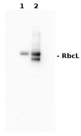
1 µg of total protein from Arabidopsis thaliana leaf (1), Hordeum vulgare leaf (2) were extracted with Protein Extraction Buffer PEB (AS08 300). Samples were diluted with 1X sample buffer (NuPAGE LDS sample buffer (Invitrogen) supplemented with 50 mM DTT and heat at 70°C for 5 min and keept on ice before loading. Protein samples were separated on 4-12% Bolt Plus gels, LDS-PAGE and blotted for 70 minutes to PVDF using tank transfer. Blots were blocked immediately following transfer in 2% blocking reagent (GE RPN 2125; Healthcare) for 1h at room temperature with agitation. Blots were incubated in the Agrisera anti-RbcL primary antibody (AS03 037) at a dilution 1: 20 000 (in blocking reagent) for 1h at room temperature. The primary antibody solution was decanted and the blot was rinsed briefly twice, and then washed 1x15 min and 3x5 min with TBS-T at room temperature with agitation. Blots were incubated in secondary antibody (goat anti-rabbit IgG horse radish peroxidase conjugated, recommended secondary antibody AS09 602, Agrisera) diluted to 1:20 000 in blocking reagent for 1h at room temperature with agitation. The blots were washed as above. The blot was developed for 5 min with AgriseraECL Bright (AS16 ECL-N-100). Images of the blots were obtained using a CCD imager (VersaDoc MP 4000) and Quantity One software (Bio-Rad). Exposure time was 5 minutes.
Note: In the barley protein sample, a well characterized 44 kDa degradation product is observed (Kokobun et al. 2002).
Caution:
Do not add any protein or HRP enzyme to reagent solution.
Optional: wash your tube, microtiter plate or membrane with 0.2 M solution of Sodium Phosphate, Dibasic, in Deionized Water. (Initial pH may be approximately 9) Bring the pH down to 8.4 with the slow addition of a 0.2M solution of Sodium Phosphate, Monobasic, in water.
25 µg of a total proteins from Arabidopsis thaliana, extracted with 50 mM Tris-HCl pH 8.5 1:1000 Triton X-100, 10 mM EDTA, 10 % Glycerol, Fresh: 1% (v/v) PIC were separated on a 10 % SDS-PAGE and transferred into nitrocellulose membrane using Iblot system. Primary antibody and secondary antibodies were used in the same dilution for both experiments.
Exposure time was:
10 minutes for a competitor's ECL
1 minute for AgriseraECLBright - Additional Information
-
Additional information: User Instruction
- Store reagents A and B in the darkness at 4-8°C.
- Mix equal volumes of reagent A and B (chemiluminescent substrate) in a clean container and equilibrate to room temperature 30 minutes before use.
- Prepare your membrane prior addition of chemiluminescent substrate, by a wash with the buffer used in your protocol (PBS or TBS or TBST-T). This will allow to remove any background prior to substrate contact.
- Optimal visualization is obtained up to 20 minutes after substrate contact. Incubation for 2-5 minutes is usually optimal.
- Remove excess substrate by filter paper.
- Cover blot with clear plastic wrap or sheet protector and expose either with x-ray film or CCD camera.
For best results clean containers and high quality water has to be used.
HS code for this product is: 3822.00.0002. - Product Citations
-
Selected references: Hao and Malnoë (2023). A Simple Sonication Method to Isolate the Chloroplast Lumen in Arabidopsis thaliana.Bio Protoc. 2023 Aug 5; 13(15): e4756.
Wieczorek et al. (2019). Contribution of Tomato torrado virus Vp26 coat protein subunit to systemic necrosis induction and virus infectivity in Solanum lycopersicum. Virol J. 2019 Jan 14;16(1):9. doi: 10.1186/s12985-019-1117-9. - Protocols
- Agrisera Western Blot protocol and video tutorials
-