Goat anti-Rabbit IgG (H&L), DyLight® 550 conjugated, min, cross-reactivity to bovine,goat,human, mouse,Rat IgG

Referentie AS111782
Formaat : 1mg
Merk : Agrisera
Neem contact op met een lokale distributeur :
 Telefoonnummer : +1 850 650 7790
Goat anti-Rabbit IgG (H&L), DyLight® 550 conjugated, min, cross-reactivity to bovine,goat,human, mouse,Rat IgG
AS11 1782 | Clonality: Polyclonal | Host:Goat | Reactivity:Rabbit IgG (H&L)
-
This product doesn't have any reviews.
| Host: | Goat |
| Clonality: | Polyclonal |
| Purity: | Immunogen affinity purified IgG. |
| Format: | Lyophilized |
| Quantity: | 1 mg |
| Reconstitution: | For reconstitution add 1,1 ml of sterile water, Let it stand 30 minutes at room temperature to dissolve, Prepare fresh working dilutions daily |
| Storage: | Store lyophilized material at 2-8°C. Product is stable for 4 weeks at 2-8°C after rehydration. For long time storage after reconstitution, dilute the antibody solution with glycerol to a final concentration of 50% glycerol and store as liquid at -20°C, to prevent loss of enzymatic activity. For example, if you have reconstituted 1 mg of antibody in 1,1 ml of sterile water add 1,1 ml of glycerol. Such solution will not freeze in -20°C, If you are using a 1:5000 dilution prior to diluting with glycerol, then you would need to use a 1:2500 dilution after adding glycerol. Prepare working dilution prior to use and then discard. Be sure to mix well but without foaming. |
| Recommended dilution: | 1 : 50 -1 : 5000 (ICC), 1 : 20 -1 : 2000 (IHC), |
Application example 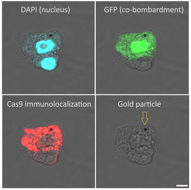 Immunolocalization of Cas9 on maize (Zea mays, cv. H1233) plant suspension cultures. Rabbit anti-Cas9 antibody (Agrisera AS16 3690), diluted 1:100 and DyLight 550 anti-rabbit secondary antibody (Agrisera AS11 1782) diluted 1:300, (red) are used for immunodetection following formaldehyde fixation, cell wall digestion and detergent permeabilization of cells. DAPI is used as nuclear marker (blue). Gold particle used for microprojectile bombardment of plasmids (GFP and Cas9) is indicated with an arrow at the last column. Scalebar is 5µm. Immunolocalization of Cas9 on maize (Zea mays, cv. H1233) plant suspension cultures. Rabbit anti-Cas9 antibody (Agrisera AS16 3690), diluted 1:100 and DyLight 550 anti-rabbit secondary antibody (Agrisera AS11 1782) diluted 1:300, (red) are used for immunodetection following formaldehyde fixation, cell wall digestion and detergent permeabilization of cells. DAPI is used as nuclear marker (blue). Gold particle used for microprojectile bombardment of plasmids (GFP and Cas9) is indicated with an arrow at the last column. Scalebar is 5µm. Courtesy of Dr. Ferhan Ayaydin, Hungarian Academy of Science, Hungary Labeling and detection protocol Fixation (30 min) 4% paraformaldehyde in PBS (pH 7.4) with 0.01% TritonX-100. (3x5min PBS wash) Cell wall digestion (40 min): 1% Cellulase, 1% Pectinase in 0.5% (w/v) MES buffer (pH 5.6) (2x5 min PBS wash) Immobilization of cells (10mins): Cells in PBS were settled onto poly-L-Lysine coated coverslips, excess PBS removed without air drying the cells. Membrane permeabilization (10 min): 0.5% TritonX-100 in PBS (3x5min PBS wash) Blocking (10mins): 5% Fish gelatin in PBS Primary antibody incubation (2.5 h at 24°C): Agrisera (AS16 3690) rabbit anti-Cas9 polyclonal antibody diluted 1:100 in blocking buffer (4x5 min blocking buffer wash) Secondary antibody incubation (1h at 24°C): goat anti-rabbit DyLight 550 antibody (Agrisera AS11 1782) diluted 1:300 in blocking buffer. (3x5min PBS wash) Nuclear counterstaining (5 min): 200ng/ml DAPI in PBS (brief PBS wash) Mounting: Fluoromount G mounting medium was used to mount coverslips onto glass slides. Imaging: Olympus FV1000 confocal microscope with 40x (NA1.3) oil immersion objective. 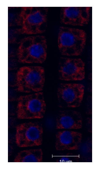 BiP localization in 4 days old Arabidopsis thaliana roots. BiP signal shown in red, Co-staining with DAPI visualized nucleus (blue color). The material has been fixed in para-formaldehyde for 30 minutes. Tissue cleaning has been performed before immunolocalization. Rabbit anti-BiP primary antibody diluted in 1: 400 and goat anti-rabbit IgG, pre-adsorbed secondary antibody DyLight®550 conjugated (Agrisera) have been used. Scale bar – 10 µm. Courtesy Dr. Taras Pasternak, Freiburg University |
| Additional information: | This antibody reacts with the heavy (gamma) chains on rabbit IgG and with the light chains on all rabbit immunoglobulins based on immunoelectrophoresis. Minimum cross-reactivity is observed to non-immunoglobulin rabbit serum proteins and bovine,goat,human,mouse,rat IgG based on immunoelectrophoresis. Antibody is supplied in 10 mM sodium phosphate, 150 mM sodium chloride, pH 7.2, 1 % (w/v) BSA, Protease/IgG free and 0.05 % (w/v) sodium azide as preservative. |
| Background: | Goat anti-rabbit IgG (H&L), DyLight® 550 conjugated, is a secondary antibody which binds to rabbit IgGs in immunological assays. DyLight® 550 has Amax = 562 nm, Emax = 576 nm. Fluor to protein ratio is 2.47 moles DyLight® 550 per mole antibody. Antibodies are adsorbed against bovine,goat,human,mouse,rat IgG and are affinity purified using solid phase rabbit IgG. DyLight® is a registered trade mark of Thermofisher Inc., and its subsidaries. |