

Polyclonal Antibody to a-internexin/NF66
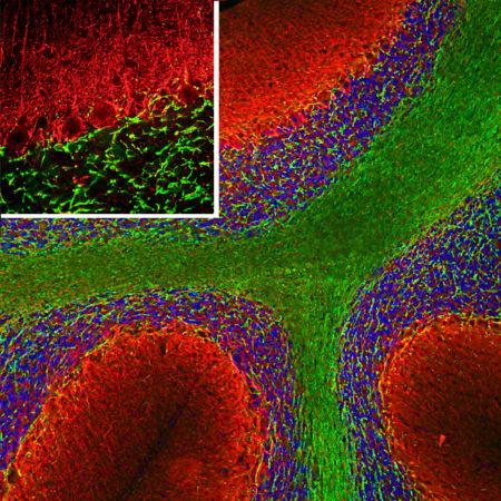
Figure-1: Immunofluorescent analysis of a rat cerebellum section stained with chicken pAb to α-internexin, (37-1054), dilution 1:5,000 in red, and costained with mouse mAb to MBP, 34-1072, 1:5,000 in green. The blue is Hoechst staining of nuclear DNA. Following transcardial perfusion of the rat with 4% paraformaldehyde, brain was post fixed for 24 hours, cut to 45μM, and free-floating sections were stained with the above antibodies. The α-internexin antibody selectively stains axons and dendrites of neuronal cells, in particular Purkinje cells, parallel fibers and the axons of granule cells, while the MBP antibody stains myelin sheathes around axons.
