

Monoclonal Antibody to Neurofilament NF-H (Clone: NAP4)
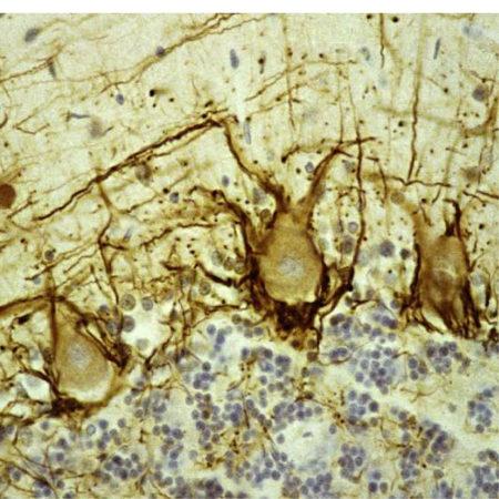
Figure-1: Immunohistological analysis of human cerebellar cortex section stained with mouse mAb to pNF-H, (34-1077), in brown. Paraffin-embedded, formalin-fixed tissue sections were stained with this antibody using the avidin biotin conjugate method. The sections was counterstained with Hematoxylin in blue. (34-1077) stains prominent basket cell axons surrounding the large Purkinje neurons. Cerebellar granule cell layer is at the bottom of the image, the molecular layer at the top.
