TrueBlot® Anti-Rabbit Ig IP Agarose Beads

Referência 00-8800-25

Tamanho : 2.5mL

Marca : Rockland Immunochemicals

Solicitar mais informações

Contactar o distribuidor local :


Telefone : +1 850 650 7790

TrueBlot® Anti-Rabbit Ig IP Agarose Beads
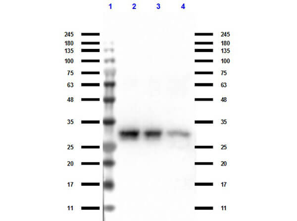
Western Blot of TrueBlot® Anti-Rabbit Ig IP Agarose Beads.
Immunoprecipitation using TrueBlot Anti-Rabbit IgG
Previous Next

Product Details

TrueBlot® Anti-Rabbit Ig IP Agarose Beads - 00-8800-25
Anti-Rabbit Immunoglobulin Gamma, Agarose-conjugated IgG, Gt-a-Rb IgG, Goat-anti-Rabbit IgG, TrueBlot for immunoprecipitation, IP beads for TrueBlot, Anti-Rabbit IgG IP agarose
Goat
Agarose ULTRA
Polyclonal
IgG
Immunoprecipitation Kit

Target Details

Rabbit
TrueBlot® Anti-Rabbit Ig IP Agarose Beads have been tested in SDS-Page, immunoprecipitation, and western blot.

Application Details

IP, SDS-PAGE, WB
ChIP, Other  - View References
Upon initial use of this product, we recommend that the vial be inverted several times to get the beads into suspension. We recommend using a large bore pipet to pipet up the liquid for use. For storage of the opened vial, we recommend that the vial cap be sealed with parafilm to help prevent evaporation of the buffer. Procedure: Preparation of Immunoprecipitated Sample for SDS-PAGE: 1. Preclear cell lysate: Add 50 μL of Anti-Rabbit IgG Beads and 500μl of cell lysate sample to a microcentrifuge tube and incubate on ice for 30 minutes. Spin at 10,000xg for 3 minutes and transfer the supernatant to a new microcentrifuge tube. 2. Immunoprecipitation: Add 5 μg of primary antibody to the microcentrifuge tube containing the precleared lysate. Incubate on ice for 1 hour. Add 50 μL of Anti-Rabbit IgG Beads. Incubate for 1 hour on a rocking platform. Spin the microcentrifuge tube at 10,000xg for 1 minute. Remove supernatant completely and wash the (pelleted) beads 3 times with 500 μL of Lysis Buffer (50mM Tris HCl, pH 8.0; 150mM NaCl; 1% NP-40). 3. Prepare sample for SDS-PAGE: After the last wash, aspirate supernatant, and add 50 μL Laemmli Buffer (with 50 mM DTT or 2% β-mercaptoethanol, final) to bead pellet. Vortex and heat to 90-100 °C for 10 minutes. Spin at 10,000xg for 3 minutes, collect supernatant, and load onto the gel. Avoid loading Anti-Rabbit Ig Beads. Note: The supernatant can be stored at -20 °C for future use. After thawing, add fresh dithiothreitol and heat as above. Centrifuge the sample at 10,000xg for 1 minute in a microcentrifuge tube to pellet any Anti-Rabbit Ig Beads and immediately transfer an aliquot of the supernatant to gel wells.

Formulation

8.9mg antibody per cc agarose
0.01 M Sodium Phosphate, 0.15 M Sodium Chloride, pH 7.2
0.09% (w/v) Sodium Azide
None

Shipping & Handling

Wet Ice
Store vial at 4 °C prior to opening. DO NOT FREEZE.
Expiration date is six (6) months from date of receipt.
TrueBlot® Anti-Rabbit Ig IP Agarose Beads are a suspension of activated agarose beads coupled with goat Anti-rabbit IgG. It is suitable for precipitation of rabbit IgGs used as the primary antibodies in immunoprecipitation assays. The beads are in suspension and will settle upon storage. Prior to use, mix the vial gently (do not vortex) to ensure delivery of proper bead volume.
(). SLC15A3 plays a crucial role in pulmonary fibrosis by regulating macrophage oxidative stress. Cell Death Differ.
Applications
IP, Co-IP
PubMed
(). Cytokine-Mediated Degradation of the Transcription Factor ERG Impacts the Pulmonary Vascular Response to Systemic Inflammatory Challenge. bioRxiv Preprint.
Applications
IP, Co-IP
PubMed
(). Deubiquitinase USP9X loss sensitizes renal cancer cells to mTOR inhibition. Int J Cancer.
Applications
IP, Co-IP
PubMed
(). Cytokine-Mediated Degradation of the Transcription Factor ERG Impacts the Pulmonary Vascular Response to Systemic Inflammatory Challenge. Arterioscler Thromb Vasc Biol.
Applications
IP, Co-IP
PubMed
(). The autophagic protein FYCO1 controls TNFRSF10/TRAIL receptor induced apoptosis and is inactivated by CASP8 (caspase 8) Autophagy
Applications
IP, Co-IP
PubMed
(). First-in-human autologous implantation of genetically modified adipocytes expressing LCAT for the treatment of familial LCAT deficiency. Heliyon.
Applications
IP, Co-IP
PubMed
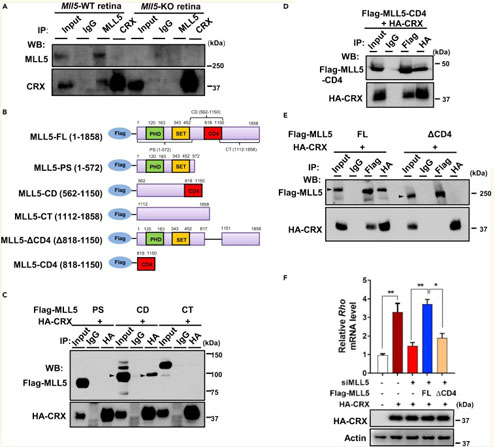
(). MLL5 is involved in retinal photoreceptor maturation through facilitating CRX-mediated photoreceptor gene transactivation. iScience.
Applications
IP, Co-IP; WB, IB, PCA
PubMed
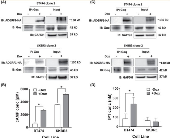
(). A novel role of ADGRF1 (GPR110) in promoting cellular quiescence and chemoresistance in human epidermal growth factor receptor 2-positive breast cancer. Faseb Journal : Official Publication of the Federation of American Societies for Experimental Biology
Applications
IP, Co-IP
PubMed
(). Upregulation of OATP1A2 in human oesophageal squamous cell carcinoma cells via the HDAC6-GCN5/PCAF-H3K9Ac axis. Xenobiotica.
Applications
IP, Co-IP
PubMed
(). Ca2+/calmodulin kinase II–dependent regulation of βIV-spectrin modulates cardiac fibroblast gene expression, proliferation, and contractility. J Biol Chem.
Applications
IP, Co-IP
PubMed
(). PFTK1 kinase regulates axogenesis during development via RhoA activation. bioRxiv Preprint
Applications
IP, Co-IP; Electronic Absorption and Fluorescence Spectroscopy
Source
(). c-Src kinase inhibits osteogenic differentiation via enhancing STAT1 stability. PLoS One
Applications
IP, Co-IP
PubMed
(). Unique Immune Cell Coactivators Specify Locus Control Region Function and Cell Stage. Mol Cell
Applications
IP, Co-IP
PubMed
(). YY1 directly interacts with myocardin to repress the triad myocardin/SRF/CArG box-mediated smooth muscle gene transcription during smooth muscle phenotypic modulation. Sci Rep.
Applications
IP, Co-IP
PubMed
(). Fermented ginseng extract, BST204, disturbs adipogenesis of mesenchymal stem cells through inhibition of S6 kinase 1 signaling. J Ginseng Res.
Applications
IP, Co-IP; WB, IB, PCA
PubMed
(). Control of cell migration by the novel protein phosphatase-2A interacting protein inka2. Cell Tissue Res.
Applications
IP, Co-IP
PubMed
(). Distinct IL-1α-responsive enhancers promote acute and coordinated changes in chromatin topology in a hierarchical manner. EMBO J.
Applications
IP, Co-IP
PubMed
(). Telomere Dysfunction Induces Sirtuin Repression that Drives Telomere-Dependent Disease. Cell Metab.
Applications
IP, Co-IP
PubMed
(). Regulation of OCT2 transcriptional repression by histone acetylation in renal cell carcinoma. Epigenetics
Applications
ChIP
PubMed
(). The interaction between SPARC and GRP78 interferes with ER stress signaling and potentiates apoptosis via PERK/eIF2α and IRE1α/XBP-1 in colorectal cancer. Nature
Applications
IP, Co-IP
PubMed
(). Single-Cell Analysis of Multiple Steps of Dynamic NF-κB Regulation in Interleukin-1α-Triggered Tumor Cells Using Proximity Ligation Assays. Cancers (Basel)
Applications
IP, Co-IP
PubMed
(). The Hippo Pathway Blocks Mammalian Retinal Müller Glial Cell Reprogramming. Cell Rep.
Applications
IP, Co-IP
PubMed
(). Two-Step Coimmunoprecipitation (TIP) Enables Efficient and Highly Selective Isolation of Native Protein Complexes. Mol Cell Proteomics
Applications
IP, Co-IP
PubMed
(). Functional role for stable microtubules in lens fiber cell elongation. Exp Cell Res
Applications
IP, Co-IP
PubMed
(). c-MET receptor as potential biomarker and target molecule for malignant testicular germ cell tumors. Oncotarget
Applications
IP, Co-IP
PubMed
(). PRDM14 directly interacts with heat shock proteins HSP90α and glucose-regulated protein 78. Cancer Science
Applications
IP, Co-IP; WB, IB, PCA
PubMed
(). GM1 ganglioside-independent intoxication by Cholera toxin. PLOS Pathogens
Applications
IP, Co-IP
PubMed
(). Inhibition of MicroRNA-155 Supports Endothelial Tight Junction Integrity Following Oxygen-Glucose Deprivation. Journal of the American Heart Association
Applications
IP, Co-IP
PubMed
(). Dermatan sulfate epimerase 1 and dermatan 4-O-sulfotransferase 1 form complexes that generate long epimerized 4-O-sulfated blocks. J Biol Chem.
Applications
IP, Co-IP
PubMed
(). c-MET receptor as potential biomarker and target molecule for malignant testicular germ cell tumors. Oncotarget
Applications
IP, Co-IP
PubMed
(). NLRP1 promotes tumor growth by enhancing inflammasome activation and suppressing apoptosis in metastatic melanoma. Oncogene
Applications
IP, Co-IP
PubMed
(). Histone deacetylase 3 prepares brown adipose tissue for acute thermogenic challenge. Nature
Applications
IP, Co-IP
PubMed
(). An HDAC3-PROX1 corepressor module acts on HNF4α to control hepatic triglycerides. Nature Communications
Applications
IP, Co-IP; WB, IB, PCA
PubMed
(). An ID2-dependent mechanism for VHL inactivation in cancer. Nature
Applications
IP, Co-IP
PubMed
(). Skin Keratins. Methods in Enzymology
Applications
IP, Co-IP
PubMed
(). BK channel inactivation gates daytime excitability in the circadian clock. Nature Communications
Applications
IP, Co-IP
PubMed
(). Leucine Carboxyl Methyltransferase 1 (LCMT-1) Methylates Protein Phosphatase 4 (PP4) and Protein Phosphatase 6 (PP6) and Differentially Regulates the Stable Formation of Different PP4 Holoenzymes. Journal of Biological Chemistry
Applications
IP, Co-IP; WB, IB, PCA
PubMed
(). Fucosylation and protein glycosylation create functional receptors for cholera toxin. Elife
Applications
IP, Co-IP
PubMed
(). Mitochondrial damage contributes to Pseudomonas aeruginosa activation of the inflammasome and is downregulated by autophagy. Autophagy
Applications
IP, Co-IP
PubMed
(). Generation of aminoterminally truncated, stable types of bioactive bovine and porcine fibroblast growth factor 4 in Escherichia coli. Biotechnol Appl Biochem.
Applications
IP, Co-IP
PubMed
(). Suppression of MAPK/JNK-MTORC1 signaling leads to premature loss of organelles and nuclei by autophagy during terminal differentiation of lens fiber cells. Autophagy
Applications
IP, Co-IP
PubMed
(). PRMT5 is upregulated in malignant and metastatic melanoma and regulates expression of MITF and p27(Kip1.) PLoS One
Applications
IP, Co-IP
PubMed
(). Early adipogenesis is regulated through USP7-mediated deubiquitination of the histone acetyltransferase TIP60. Nat Commun.
Applications
IP, Co-IP
PubMed
(). c-Jun N-terminal kinase phosphorylates DCP1a to control formation of P bodies. J Cell Biol.
Applications
IP, Co-IP
PubMed
(). Phosphorylation of profilin by ROCK1 regulates polyglutamine aggregation. Mol Cell Biol
Applications
IP, Co-IP
PubMed
(). c-Jun controls histone modifications, NF-kappaB recruitment, and RNA polymerase II function to activate the ccl2 gene. Mol Cell Biol. 2008;28(13):4407-4423. doi:10.1128/MCB.00535-07 Mol Cell Biol.
Applications
IP, Co-IP
PubMed
(). Selective cochlear degeneration in mice lacking the F-box protein, Fbx2, a glycoprotein-specific ubiquitin ligase subunit. J Neurosci.
Applications
IP, Co-IP
PubMed

What is Rockland’s TrueBlot® product line?

Rockland’s TrueBlot® product line is designed to solve common experimental problems when performing immunoprecipitation/co-immunoprecipitation experiments and Western blots of immunoprecipitated samples (IP–Western blot). The product line consists of TrueBlot® monoclonal secondary antibodies and TrueBlot® IP beads.

What is the advantage of TrueBlot® secondaries over regular secondary antibodies?

TrueBlot® antibodies are specific to the whole IgG molecule and will not bind heavy or light chains. This is useful for binding target proteins with an expected MW near 25 kDa (light chains) and 50 kDa (heavy chains).

Why does it appear that the TrueBlot® antibody is binding heavy/light chains in my sample?

The sample must be fully reduced to eliminate cross-reactivity with heavy and light chains. Any reactivity to heavy and light chains could be due to incomplete reduction of your sample. Please be sure to optimize reducing conditions for your sample type.

How can I ensure my samples are fully reduced?

Samples can be fully reduced by heating at 90-100°C for 10 minutes in sample buffer containing reducing agent (for example, DTT to a final concentration of 50 mM or add β-Mercaptoethanol to a final concentration of 2% (v/v)).

With which species/isotypes are TrueBlot® secondary antibodies reactive?

TrueBlot® secondary antibodies are reactive with the IgG of their respective species. For example, if using a primary antibody raised in mouse (mouse host), use TrueBlot® anti-mouse Ig secondary antibodies for detection.

Do TrueBlot® secondary antibodies detect IgM?

TrueBlot® secondary antibodies have not been shown to detect IgM.

Do I need to preclear my lysate before the immunoprecipitation step?

Samples that have many other proteins present (such as lysates) may require preclearing to prevent interference in later IP and WB procedures. Recombinant protein samples require pre-clearing more often than serum samples.

How can I enrich/concentrate my lysate for a low expression of a protein of interest?

The immunoprecipitation procedure can be repeated several times to yield a more concentrated protein solution.

What is the recommended lysis buffer for TrueBlot® products?

The optimal lysis buffer will depend on your sample type. See protocol for buffer recommendations. Generally, 1X RIPA is used to lyse samples.

My target protein has the same MW as a heavy/light chain. How can I be sure that the band is the target protein and not a heavy/light chain?

Be sure to include a positive control for the primary antibody in your experiment and check that the sample is fully reduced.

What is the average size of TrueBlot® magnetic/agarose beads?

The beads are roughly 0.5 μm in diameter.

Why can’t I use Protein A or Protein G beads for the IP step in IP–Western blot when using rabbit species?

Using Protein A or Protein G beads with rabbit species results in contaminated bands. For best results when using rabbit species, use Rockland’s TrueBlot® Rabbit Ig IP beads, which are available in either a magnetic or agarose format. Please see individual TrueBlot® Rabbit product pages for additional details.

Can I use Protein A or Protein G beads for the IP step in IP–Western blot when using mouse, goat, or sheep species?

Yes. If you are using Protein A or Protein G beads with mouse, goat, or sheep species, we recommend the following: Determine the compatibility of Protein A or Protein G with your species and subtype, do not use excessive amounts of slurry, and choose the appropriate elution conditions. In some instances, harsher elution conditions can cause stripping of the subunits of Protein A or Protein G from the bead support and result in non-specific bands. For best results, we recommend using TrueBlot® Ig IP beads, which are available in either agarose or magnetic format.

This product is for research use only and is not intended for therapeutic or diagnostic applications. Please contact a technical service representative for more information. All products of animal origin manufactured by Rockland Immunochemicals are derived from starting materials of North American origin. Collection was performed in United States Department of Agriculture (USDA) inspected facilities and all materials have been inspected and certified to be free of disease and suitable for exportation. All properties listed are typical characteristics and are not specifications. All suggestions and data are offered in good faith but without guarantee as conditions and methods of use of our products are beyond our control. All claims must be made within 30 days following the date of delivery. The prospective user must determine the suitability of our materials before adopting them on a commercial scale. Suggested uses of our products are not recommendations to use our products in violation of any patent or as a license under any patent of Rockland Immunochemicals, Inc. If you require a commercial license to use this material and do not have one, then return this material, unopened to: Rockland Inc., P.O. BOX 5199, Limerick, Pennsylvania, USA.

Related Resources

Você também pode estar interessado nos seguintes produtos:


Referência
Descrição
Cond.
Price Bef. VAT
ARP59155_P050
 100ul