
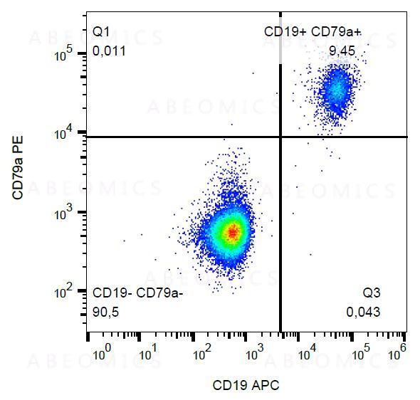
Figure 1: Intracellular staining of CD79a in human peripheral blood with anti-CD79a (HM57) PE.
Tamanho : 100Testss
 Telefone : +1 850 650 7790
View all pathways
View all interactive pathways
Figure 1: Intracellular staining of CD79a in human peripheral blood with anti-CD79a (HM57) PE.
| Amount : | 100 tests |
| Isotype : | Mouse IgG1 |
| Storage condition : | Store in the dark at 2-8°C. Do not freeze. Avoid prolonged exposure to light. |
| Subcellular location: | Cell membrane |
| Post transnational modification: | Arginine methylation in the ITAM domain may interfere with the binding of SYK. It promotes signals leading to B-cell differentiation (By similarity). |
| Tissue Specificity: | B-cells. |
| BioGrid: | 107411. 53 interactions. |
| There are currently no product reviews |
